
Overview of Pediatric Atlases Release V1.1:

Release Date: 08/05/2013 | Version 1.0
Three mean atlases dedicated for pediatric age clusters: 0 – 2 y.o., 2 – 9 y.o, and 9 – 18 y.o are available to download in Release V1.1.

Components of downloadable archived file bundles for each atlas:

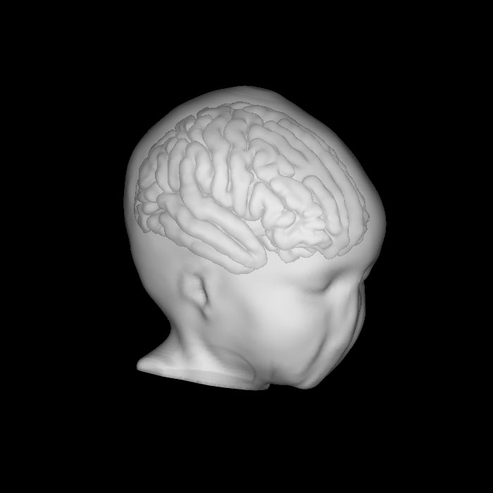
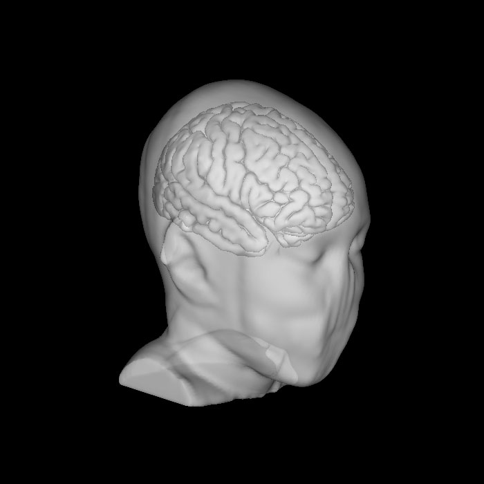
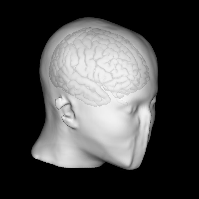
  • Defaced high resolution (1 mm^3)  segmented MRI/CT co-registered volume (7 different tissues are labelled:  external air, scalp, skull, CSF, GM, WM and internal air)
  • Separate file with dipole coordinates on cortex;
  • Separate files of generic 128/256 electrodes montages, co-registered to each atlas (in the same xyz space)
  • Lead Field Matrices (LFM) for EGI Geodesic generic 128/256/Super Dense electrodes montages
  • Generic (super dense) LFM to use in any montage by interpolation
  • Matlab scripts for uploading binaries into Matlab
  • ReadMe file (with Metadata, file formats, and other technical details).

Authorization Required to Download Atlases

Before downloading the atlases, you must complete and sign a Data Use Agreement to obtain access privileges to the restricted downloads. If you have already been authorized, login to access the data.

Please include your user name (not password) in the Data Use Agreement.  Please also include in the Data Use Agreement your academic or corporate affiliation. Once you complete the Data Use Agreement, you will be contacted by return e-mail when access privileges are in place.  At that point, upon logging in, you may download the head models.

The purpose of the data use agreement is to comply with the requirements of our Institutional Review Boards and to avoid infringement of existing US Patents.  This is spelled out in the terms of the data use agreement.

Atlas 1 (0-2 years old)

0-2-lrg

Atlas 2 (2-8 years old)

4-8-lrg

Atlas 3 (9-18 years old)

8-18-lrg

Downloads

Methods:

The atlases were created by fusion (nonlinear warping) of a publicly available probabilistic UNC non-linear- average MRI template for 1 y.o. (Shi et al, 2011),  MNI non-linear- average MRI template for  4-8 y.o., and MNI nonlinear average MRI template for 10-14 y.o. (Fonov et al, 2011) to our age matched CTs, and subsequent segmentation with BrainK. The quality of segmentation was validated by visual slice-by-slice inspection and manual correction. The quality of nonlinear warping of brain templates into CT crania was validated by matching the CSF layer thickness to the average CSF developmental curves at several cranium landmarks.  Lead Field Matrices were computed on the University of Oregon ACISS cluster using a Finite Difference Model (FDM) solver (Song et al,  2013). The following conductivity values were used based on the average literature data: 0.46 S/m (scalp), 0.018 S/m (skull), 1.79 S/m (CSF), 0.25 S/m (GM), 0.33 S/m (WM) and 0 S/m ( air). Quality of the LFMs was validated against head models generated from subject specific CT/MRI.

Works Cited:

Song J, Morgan K, Turovets S, LI K, Davey C, Govyadinov P, Luu P, Smith K, Prior F, Larson-Prior L, Tucker D. Anatomically Accurate Head Models and Their Derivatives for Dense Array EEG Source Localization. Funct Neurol Rehabil Ergon 2013; 3(2-3):275-293. (Paper)